


To investigate the handling and electronic programmability of lipid containers, Giant Unilamellar Vesicles (GUVs) were chosen as model system. They can be detected readily by Differential Interference Contrast (DIC) or fluorescence microscopy. Furthermore, various methods for formation of GUVs are in common use [1]. Only marginal interactions with the hydrophobic channel walls were found, so that extremely slow flow rates in PDMS-microchannels can be achieved (<1µl/h). We then demonstrated vesicle manipulation (retention and release) by digitally pulsed fields (opposite figure, with strong effects because of a relatively high dielectric-constant, eliminating the need for charged lipid membranes. [2]
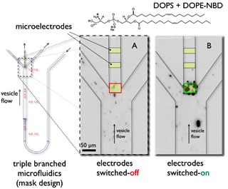
The inverted fluorescence images showing electronic manipulation of GUVs (DOPS: 1,2-Dioleoyl-sn- Glycero-3[Phospho-L-Serine] labelled by NBD: 7-nitrobenz-2-oxa-1,3-diazole-4-yl) in a triple branched microfluidic channel geometry. The electrodes were visualized by coloured rectangles inside the middle channel. There are two states: image A. electrodes switched-off – the main liposome material flows through the outer channel arms, and image B. electrodes switched-on with liposome accumulation on the upper electrode and reinforced material-flow in the middle channel.
The shown experiment performed with individually programmable microelectrodes has demonstrated the possibility to concentrate and route GUVs by digitally pulsed fields in microfluidic geometries.
REFERENCES
[1] P. L. Luisi, P. Walde (eds.) in Giant Vesicles, John Wiley & Sons, 1999.
[2] P. F. Wagler, U. Tangen, M. Heymann, T. Maeke, S. Chemnitz, M. Jünger, T. Palutke, J. S. McCaskill, in µTAS 2005, Boston, 2005, pp. 1134-1136.